 4.9 Rating | 23.000+ Procedures | Medical Experts

Table of contents:

What problems can a dental check-up identify?
Dental examination: what does it assess and how long does it take?
Panoramic dental X-ray: what does it show and when is it needed?
3D intraoral scanner: how does it work and what is it used for?
How are the diagnosis and treatment plan defined?
How often should you have a dental check-up?
Why choose KEIT Dental for dental check-ups and diagnosis?
In this article, you will learn which problems a dental check-up can detect, what is assessed during the examination, and when panoramic dental X-rays, intraoral X-rays, CBCT scans, and 3D intraoral scanning may be recommended. We also explain how a treatment plan is developed, how often dental check-ups should be scheduled, and why you should choose KEIT Dental.
 Book your FREEOnline Consultation Now

What problems can a dental check-up identify?
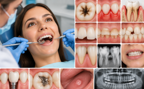
A dental check-up can detect tooth decay, gum and periodontal problems, infections, fractures, tooth wear, malocclusions, and abnormalities of the oral tissues, even before noticeable symptoms appear. During the examination, the dentist also assesses existing fillings, dental restorations, prostheses, implants, and other previous treatments, checking for signs of leakage, wear, or deterioration.
The dentist also examines the oral mucosa, including the tongue, cheeks, palate, and floor of the mouth, to identify any lesions, spots, or abnormalities that require monitoring or further investigation. When a clinical examination alone is insufficient, the assessment may be supplemented with X-rays or digital diagnostic tools to examine structures and details that are not directly visible.
Dental examination: what does it assess and how long does it take?
A dental examination assesses the teeth, gums, oral mucosa, bite and existing restorations and usually takes approximately 20–30 minutes, unless additional examinations or more detailed diagnostic investigations are required. The dental consultation begins with a medical and dental history, during which the dentist collects information about current symptoms, previous medical conditions, allergies, medications and earlier dental treatments. During a first consultation, the patient’s functional and aesthetic needs are also evaluated.
This is followed by a clinical examination of the oral cavity. The dentist checks the teeth for decay, fractures and wear, assesses the gums for inflammation or bleeding and examines the oral mucosa for any abnormalities. The bite is also evaluated to identify chewing difficulties, tooth misalignment or problems affecting the relationship between the upper and lower dental arches.
When clinically necessary, the dental examination may include specific tests such as pulp vitality testing, assessment of tooth mobility, measurement of periodontal pocket depth and evaluation of jaw function. A routine follow-up appointment may be shorter, while the presence of symptoms or the need for diagnostic imaging may extend the duration of the visit. When a complete view of the teeth and jaw bones is required, the dentist may recommend a panoramic dental X-ray or other targeted radiographs to support a more accurate diagnosis.
Panoramic dental X-ray: what does it show and when is it needed?
A panoramic dental X-ray shows the teeth, dental arches and jaw bones in a single image and is recommended when the dental examination requires an overall assessment of structures that cannot be seen directly. Also known as an orthopantomogram or OPG, it may reveal impacted teeth, wisdom teeth, bone loss, root abnormalities and possible lesions affecting the jaws. A panoramic dental X-ray is commonly used during the initial assessment for orthodontic, implant or oral surgery treatment and whenever both dental arches need to be examined together. The procedure is quick and painless. The machine rotates around the patient’s head while the patient remains still. Before the image is taken, metal objects that could interfere with image quality must be removed.
A panoramic X-ray provides a general two-dimensional view, but it does not show every detail of an individual tooth or the three-dimensional thickness of the bone. When more specific information is needed, the dentist may request intraoral radiographs, CBCT imaging or another targeted examination. A 3D intraoral scanner is used instead when the aim is to record the shape and position of the visible dental surfaces digitally.
3D intraoral scanner: how does it work and what is it used for?
A 3D intraoral scanner captures optical images of the teeth and gums and creates a digital impression that can be used to design restorations, prostheses and orthodontic treatments. During the scan, the dentist moves a small handheld device inside the mouth. The software processes the images in real time and reconstructs a three-dimensional model of the dental arches, which can be reviewed immediately and sent digitally to the dental laboratory.
The intraoral scanner is used when planning veneers, crowns, bridges, dental prostheses, night guards and clear aligners. Compared with conventional impressions, it avoids the use of impression materials and may improve patient comfort, particularly in people with a pronounced gag reflex. The scan does not use X-rays and records only the visible surfaces of the teeth and gums. It therefore does not show the tooth roots, bone or internal anatomical structures and cannot replace radiographic examinations when these are clinically necessary. Panoramic radiographs, intraoral radiographs, CBCT scans and intraoral scanning provide different types of diagnostic information and are selected according to the specific problem that needs to be assessed.
How are the diagnosis and treatment plan defined?
A dental diagnosis is established through a structured process that combines a complete medical and dental history, a detailed clinical examination and targeted diagnostic investigations such as dental radiographs, panoramic X-rays or dental CT scans. The dentist considers the symptoms reported by the patient, evaluates the condition of the teeth, gums and oral tissues and identifies any conditions such as tooth decay, infection or periodontal disease. Based on this information, a personalized dental treatment plan is prepared. The plan defines the treatments required, the order of clinical priorities, the expected treatment schedule and the overall costs, allowing care to be organised according to the patient’s oral health needs.
The dental treatment plan includes the sequence of procedures, the expected number of appointments, possible treatment alternatives and the risks associated with each intervention. More urgent conditions, such as active infections or gum inflammation, are treated before restorative, rehabilitative or cosmetic procedures so that treatment begins from a stable clinical foundation. Although photographs and radiographs sent remotely may support an initial assessment, a definitive diagnosis always requires an in-person examination at the dental clinic. After treatment has been completed, the dentist establishes a schedule of follow-up appointments and professional oral hygiene sessions based on the patient’s individual level of risk, helping to maintain the results over time.
How often should you have a dental check-up?
A dental check-up is generally recommended every 6–12 months, although the appropriate frequency depends on the patient’s individual risk factors and the condition of the teeth and gums. More frequent examinations may be required for patients with recurring tooth decay, periodontal disease, dental implants, prostheses, orthodontic appliances, diabetes, smoking habits or inadequate oral hygiene.
The dental appointment should be brought forward if symptoms such as pain, swelling, persistent gum bleeding, tooth mobility, dental fractures or difficulty chewing occur. Dental radiographs are not automatically taken at every check-up. They are performed only when they are clinically necessary, according to the symptoms, previous findings and current oral health status.
The recommended frequency of dental appointments and the need for any diagnostic investigations may also influence the overall cost of monitoring and treatment.
Why choose KEIT Dental for dental check-ups and diagnosis?
KEIT Dental is the dental clinic of the KEIT brand, integrated into the KEIT Day Hospital healthcare facility in Tirana, Albania, founded in 2011 by Dr Skerdilajd Faria. With over 23,000 procedures performed across the KEIT brand's various areas of activity, the facility combines specialist expertise, medical technology, safety protocols, and personalised patient care.
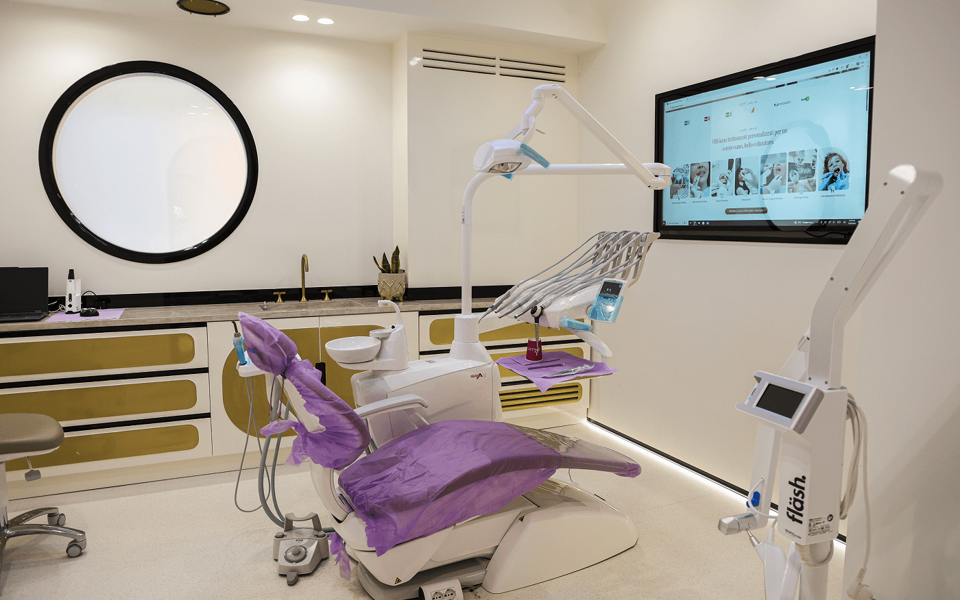
KEIT Dental has seven dental treatment rooms and an in-house dental laboratory. Its integration with KEIT Day Hospital also provides access to a multidisciplinary medical team, oral and maxillofacial surgeons, specialists in anaesthesia and intensive care, equipped operating theatres, and inpatient wards for cases requiring more complex care. For dental check-ups and diagnosis, KEIT Dental adopts a personalised approach aimed at identifying potential conditions early, assessing oral health, and developing an appropriate treatment plan based on the patient's needs.
1. Seven dental treatment rooms and diagnostic technologies
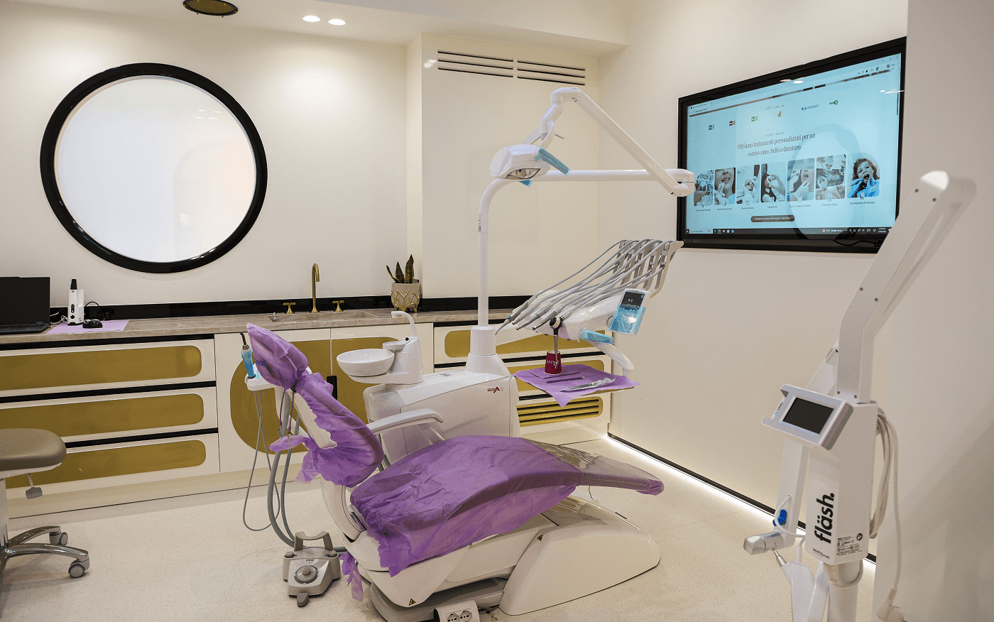
KEIT Dental has seven treatment rooms dedicated to dental care, organised to accommodate restorative dentistry, endodontics, implantology, and prosthetic rehabilitation procedures. During...
KEIT Dental has seven treatment rooms dedicated to dental care, organised to accommodate restorative dentistry, endodontics, implantology, and prosthetic rehabilitation procedures. During dental check-ups, diagnostic instruments and imaging technologies, such as intraoral X-rays, panoramic dental X-rays, and 3D intraoral scanners, allow a more detailed assessment of the teeth, gums, bite, and oral structures. When clinically indicated, CBCT scans also provide three-dimensional images of bone structures. Diagnostic examinations are selected according to clinical needs, avoiding unnecessary investigations.

2. Multidisciplinary dental and medical team
One of the main advantages of KEIT Dental is its ability to combine dental expertise with the multidisciplinary medical team at KEIT...
One of the main advantages of KEIT Dental is its ability to combine dental expertise with the multidisciplinary medical team at KEIT Day Hospital. The clinic works with professionals experienced in restorative dentistry, endodontics, implantology, and dental prosthetics. In particularly complex cases, oral and maxillofacial surgeons and other medical specialists may also be involved, depending on clinical needs.
When diagnosing complex dental conditions, collaboration between professionals allows the assessment of teeth, gums, bone structures, and occlusion to be coordinated, helping identify any need for specialist treatment. This approach supports the development of a coordinated treatment plan, including cases requiring procedures from different dental specialties.
3. Thorough diagnosis and personalised medical assessment
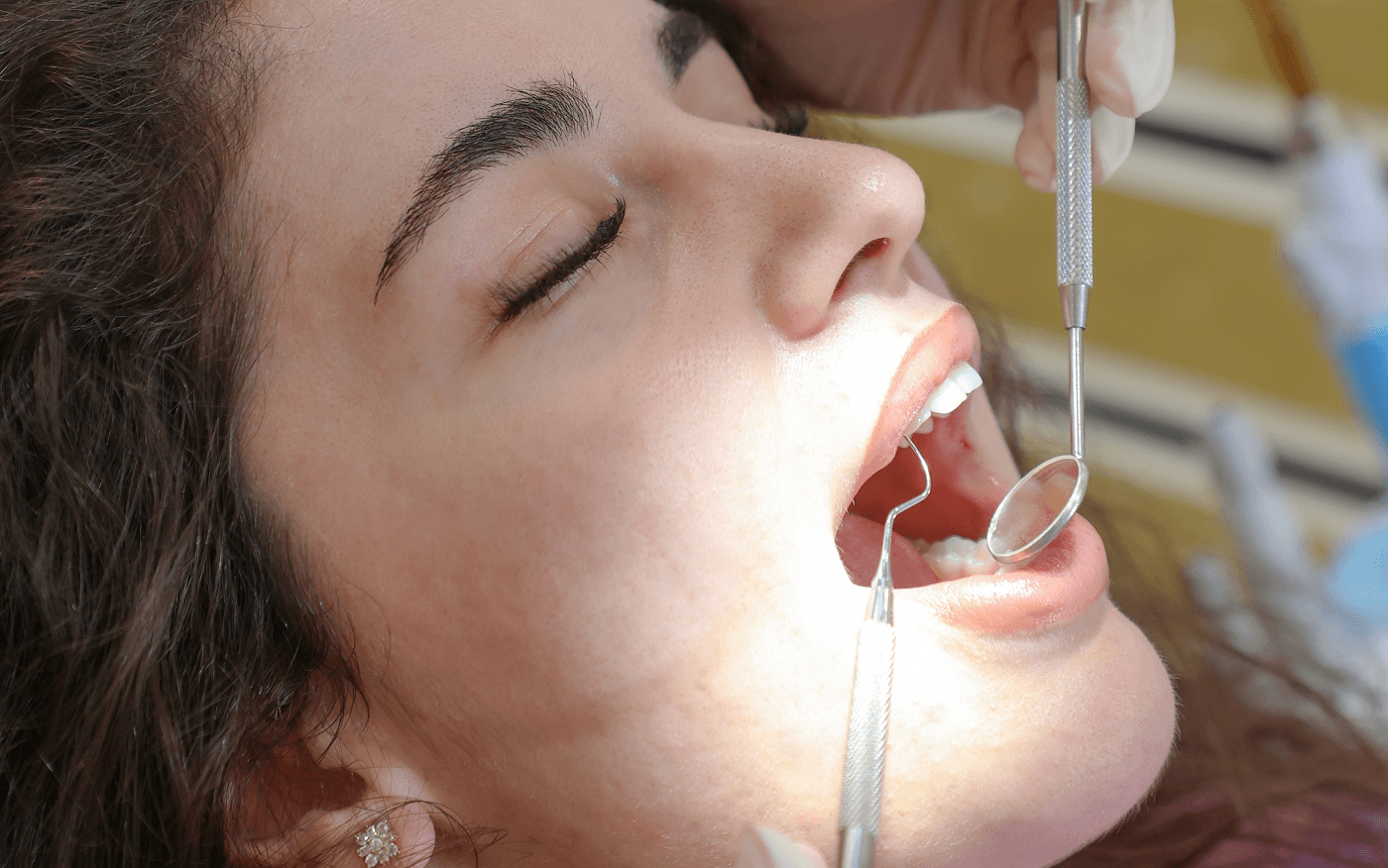
During the appointment, the dentist reviews the patient's medical history and assesses the condition of the teeth, gums, oral mucosa, and bite,...
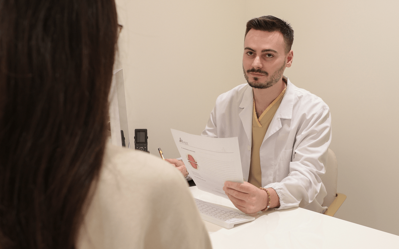
During the appointment, the dentist reviews the patient's medical history and assesses the condition of the teeth, gums, oral mucosa, and bite, also checking existing restorations, prostheses, and implants. When indicated, radiographic examinations and digital scans provide additional information to support the clinical assessment. Particular attention is paid to identifying tooth decay, infections, periodontal diseases, and abnormalities of the oral structures, distinguishing conditions requiring prompt intervention from those that can be monitored.
Thanks to its integration with KEIT Day Hospital, patients with particular medical conditions or those requiring more complex procedures may undergo assessments by anaesthetists, cardiologists, or other specialists, together with any necessary diagnostic evaluations. Treatment planning therefore considers both the patient's dental needs and general health.
4. In-house dental laboratory and digital restoration planning
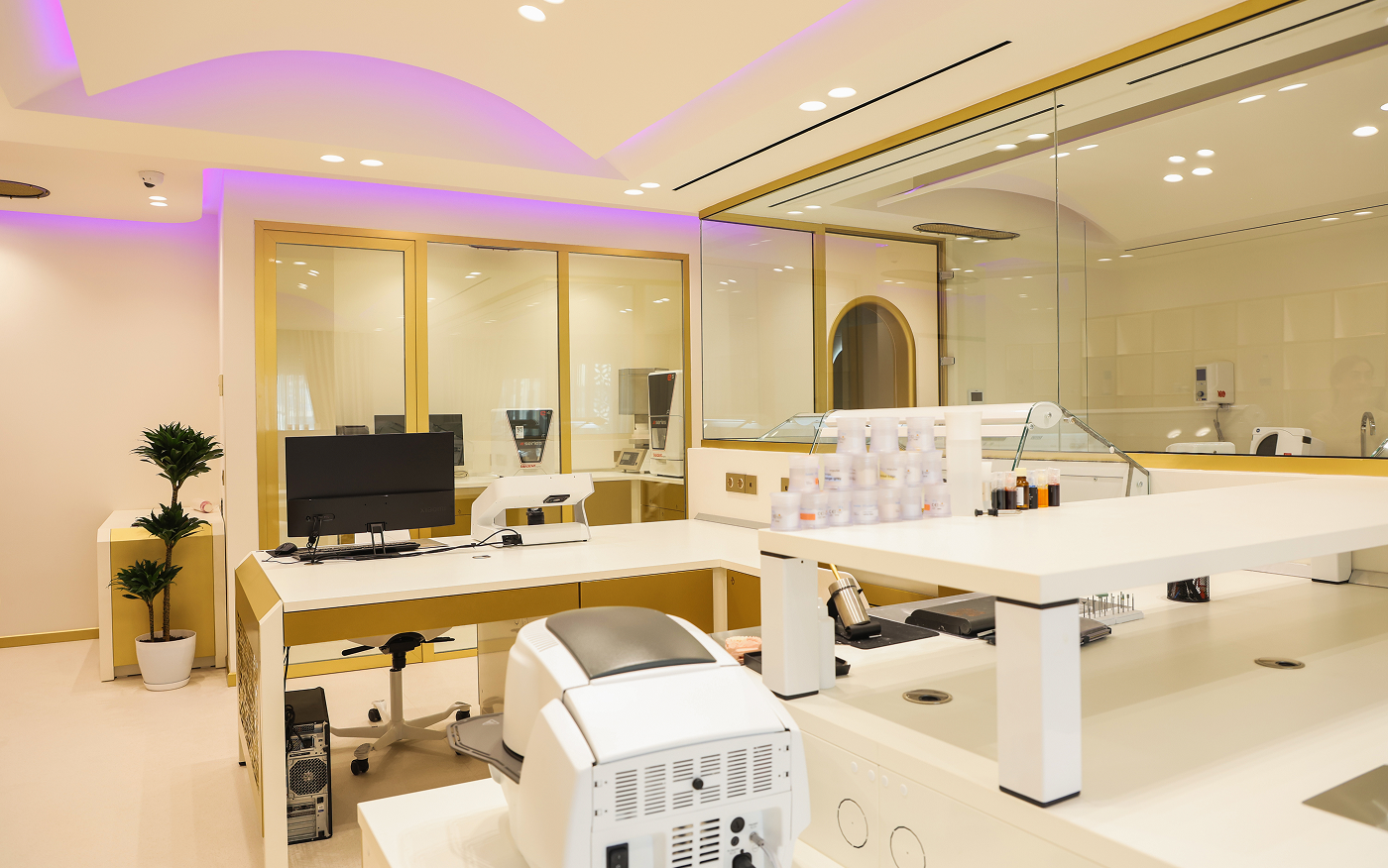
One of KEIT Dental's distinguishing features is its in-house dental laboratory, which facilitates direct collaboration between dentists and dental technicians in producing...
One of KEIT Dental's distinguishing features is its in-house dental laboratory, which facilitates direct collaboration between dentists and dental technicians in producing restorations and prosthetic rehabilitations. When a diagnostic assessment identifies the need for crowns, bridges, or other restorations, digital impressions obtained using a 3D intraoral scanner can be used to design customised restorations according to their shape, dimensions, and occlusal relationships.
Coordination between the clinic and laboratory allows clinical data and technical details to be shared, with particular attention to design precision, chewing function, and the aesthetic harmony of the final result.
5. Safety, sterilisation, and access to hospital infrastructure
KEIT Dental follows hygiene and infection control protocols that include disinfecting treatment areas, sterilising instruments, and using disposable materials when indicated. During...
KEIT Dental follows hygiene and infection control protocols that include disinfecting treatment areas, sterilising instruments, and using disposable materials when indicated. During diagnostic procedures, particular attention is paid to the proper handling of instruments and ensuring that radiographic examinations are performed only when justified by clinical needs.
For cases requiring prolonged monitoring or specific postoperative care, admission to KEIT Day Hospital's inpatient wards can also be arranged. These wards are equipped with healthcare staff and monitoring systems. This integration allows treatment to be organised according to the level of care required by the patient's condition and the complexity of the procedure.
6. Personalised care and regular dental check-ups
At KEIT Dental, patient care continues after the diagnostic appointment through a personalised treatment plan and follow-up appointments scheduled according to clinical...
At KEIT Dental, patient care continues after the diagnostic appointment through a personalised treatment plan and follow-up appointments scheduled according to clinical needs. Patients receive guidance on any necessary treatments, treatment priorities, and preventive measures to maintain oral health.
Subsequent appointments allow the dentist to monitor the condition of the teeth, gums, and restorations, identifying any changes at an early stage. Dental check-ups are generally scheduled every 6–12 months, with shorter intervals for patients at higher risk. In complex cases, care may also involve other specialists from the KEIT team. For international patients, care may continue after they return to their home country through communication with the clinic and guidance on necessary follow-up appointments.

7. Transparency, clinical documentation, and guarantee conditions
KEIT Dental places particular emphasis on transparency throughout the diagnostic process and clinical documentation. After the examination, patients receive information about the...
KEIT Dental places particular emphasis on transparency throughout the diagnostic process and clinical documentation. After the examination, patients receive information about the conditions identified, any additional examinations required, available treatment options, expected benefits, and potential risks.
Clinical documentation includes diagnostic examinations, the assessments performed, and the proposed treatment plan. The informed consent form, when required, and treatment quotation also allow patients to understand the details of the examinations and any proposed treatments, together with their associated costs. Any applicable guarantee conditions relating to treatment procedures are specified in the contractual documentation, clarifying the coverage provided, exclusions, and arrangements for assistance.
FAQ
Frequently asked questions
Can I have a dental check-up even if I have no pain?

Yes. Dental check-ups should not be scheduled only when pain or other symptoms appear. Early tooth decay, gum problems, tooth wear, changes in dental restorations, and certain oral conditions can develop without causing noticeable symptoms. Regular dental check-ups therefore help detect potential problems in their early stages.
Do I need to bring previous dental X-rays to my appointment?
Yes, if available. Previous dental X-rays can provide useful information and, if they are sufficiently recent and of good quality, may help avoid repeating unnecessary examinations. The dentist will assess whether the available images are suitable for evaluating the problem.
Can a panoramic dental X-ray replace an intraoral X-ray?
No. A panoramic dental X-ray provides an overall view of the dental arches and surrounding structures, while an intraoral X-ray allows a more detailed examination of one or a few teeth. The choice therefore depends on the area being examined and the information required for diagnosis.
Can a 3D intraoral scanner detect tooth decay?
It is not the primary tool for diagnosing tooth decay. An intraoral scanner captures the visible surfaces of the teeth and gums and can be useful for digital assessment and treatment planning, but it does not show the internal structures of the teeth. When necessary, diagnosis is supplemented with a clinical examination and targeted dental X-rays.
Is a CBCT scan always more accurate than a panoramic dental X-ray?
CBCT provides more detailed three-dimensional images, but this does not mean it is always the most appropriate examination. A panoramic dental X-ray may be sufficient for a general assessment, while CBCT is prescribed when specific three-dimensional information about the teeth, bone, or anatomical structures is required. The choice should therefore be based on clinical necessity.