Published on:
 Modified on:

 Reading Time: 4 min
 Aesthetic Treatments
Xanthelasma is an eyelid condition in which yellowish plaques develop due to the deposition of lipids in the skin. It may occur in association with lipid abnormalities, although it can also be seen in individuals with normal cholesterol levels.
 Table of Contents:
-
1
 Xanthelasma: What it is and where it appears
-
2
What are the symptoms and appearance of xanthelasma?
-
3
Xanthelasma causes: What factors are associated with it?
-
4
Xanthelasma: How it is diagnosed
-
5
Xanthelasma treatment and removal options
-
6
Xanthelasma removal cream and home treatments: Do they work?
-
7
What to expect after xanthelasma removal: Healing and aftercare
-
8
Can xanthelasma come back after removal?
In this guide, we explain what xanthelasma is and where it appears, its symptoms, what causes it and how it is diagnosed. We also review the available treatment and removal options, healing and aftercare after removal, and the possibility of recurrence over time.
Xanthelasma: What it is and where it appears
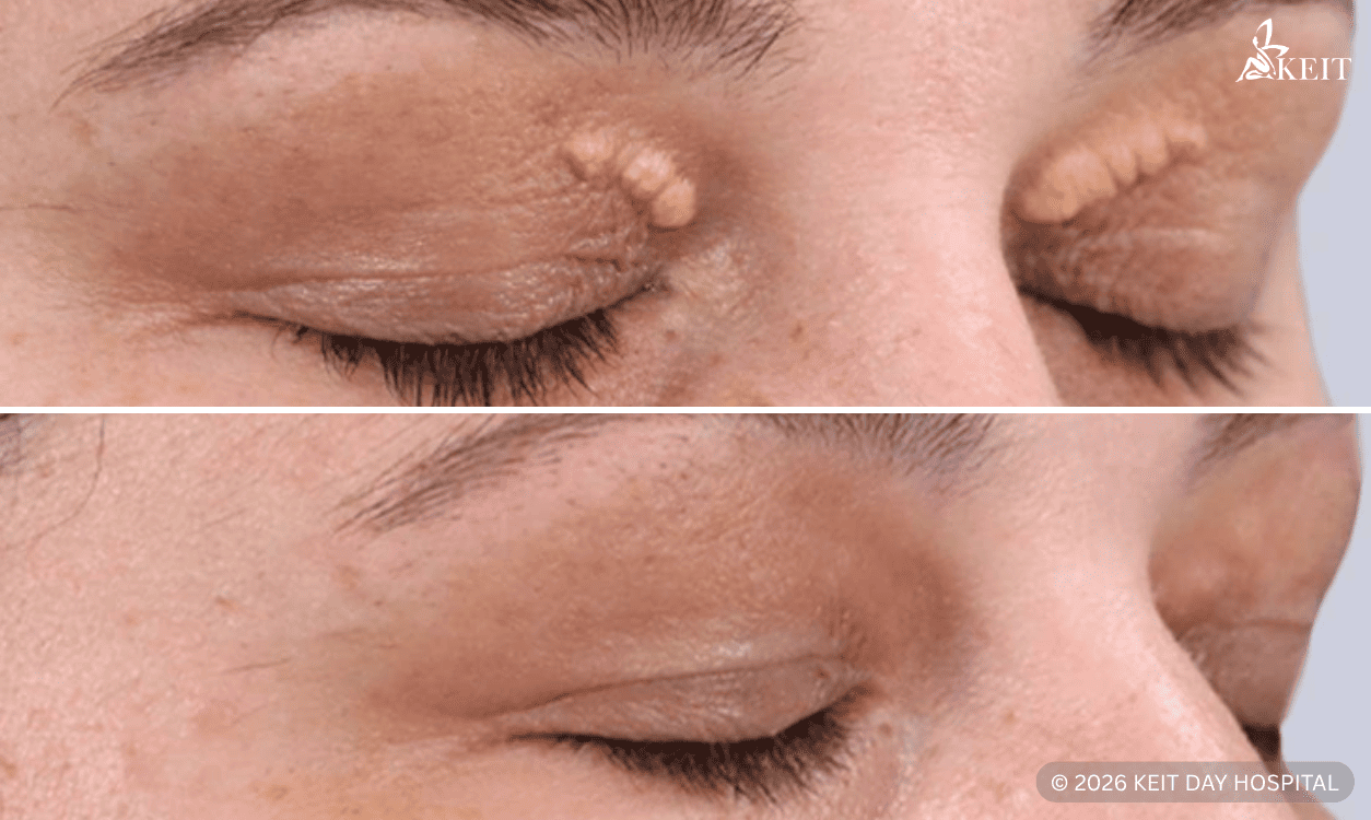
Xanthelasma is a benign condition characterized by the accumulation of lipid-rich deposits within the superficial layers of the skin, most commonly on the eyelids. It typically appears as soft, yellowish plaques and most often develops near the inner corners of the upper eyelids, although the lower eyelids may also be affected. In many cases, the lesions are bilateral and symmetrically distributed.
What are the symptoms and appearance of xanthelasma?
Xanthelasma is mainly identified by its eyelid appearance rather than physical symptoms. Common features include:
- Yellow or yellow-white plaques on the upper or lower eyelids.
- Flat or slightly raised lesions with a smooth surface.
- Symmetrical distribution, often near the inner corners of the eyelids.
- Gradual enlargement over time.
- Usually painless and not associated with inflammation.
Xanthelasma causes: What factors are associated with it?
Xanthelasma is mainly associated with lipid accumulation in the skin, often related to underlying metabolic or cholesterol-related factors. The main contributing factors include:
- Abnormal lipid levels: High cholesterol or other lipid disorders are commonly associated with xanthelasma.
- Genetic susceptibility: Inherited dyslipidaemias, including familial hypercholesterolaemia, can increase the likelihood of cholesterol-rich skin deposits.
- Associated conditions: Diabetes, thyroid disease, and other metabolic conditions may coexist with xanthelasma.
- Normal lipid profile in some patients: Xanthelasma can still occur even when cholesterol levels are within the normal range.
Xanthelasma: How it is diagnosed
Xanthelasma is diagnosed clinically, based on its typical appearance and location on the eyelids. In most cases, the doctor evaluates the following:
- Appearance of the lesion: Its color, surface, borders, and whether it is flat or slightly raised.
- Location and distribution: Whether it affects the upper or lower eyelids, one side or both, and the area near the inner canthus.
- Lipid profile, when indicated: Blood tests may be requested to assess cholesterol and other lipid abnormalities.
- Differential diagnosis: Other eyelid lesions may be considered if the appearance is atypical or the diagnosis is uncertain.
Xanthelasma treatment and removal options
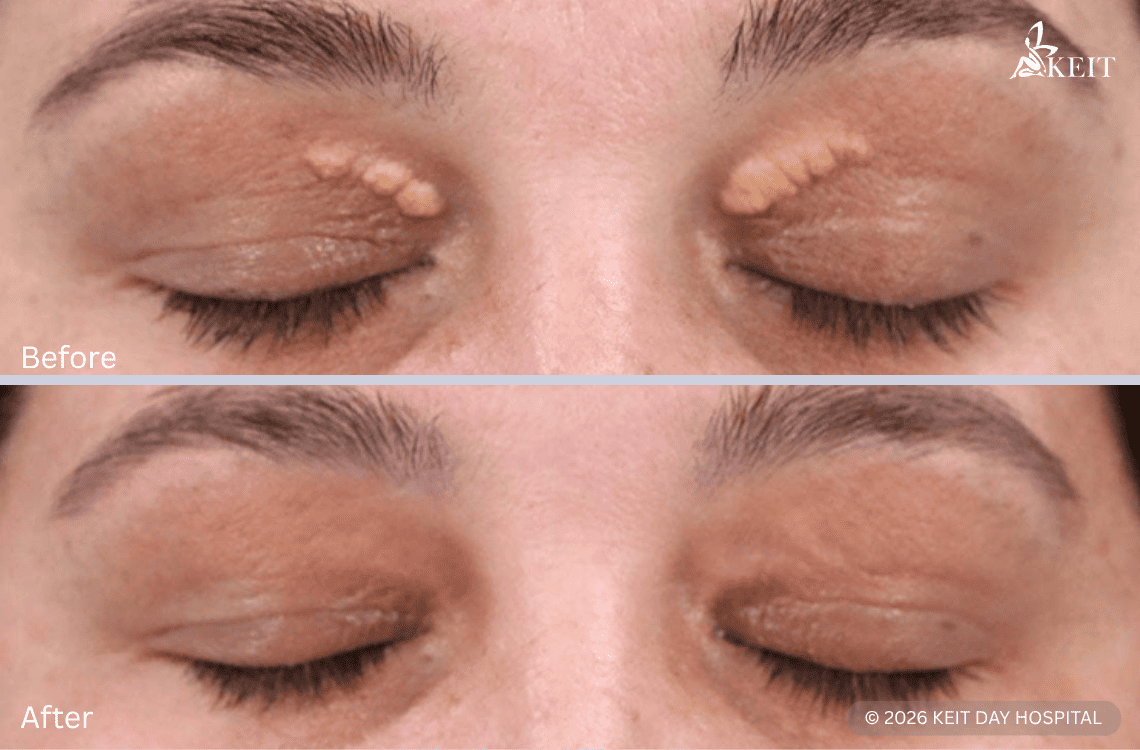
Xanthelasma treatment is selected according to the size, depth, location, and extent of the lesions, as well as the expected cosmetic outcome. The main xanthelasma removal options include:
- Surgical excision: Removal of the plaque with careful eyelid technique, usually considered for selected lesions.
- Laser treatment: Various lasers may be used to treat xanthelasmas, particularly for cosmetic removal.
- Chemical peeling: Uses controlled application of chemical agents, such as trichloroacetic acid (TCA), to treat superficial xanthelasma plaques.
- Plasma arc treatment: Uses controlled plasma energy to ablate superficial lesions and promote skin remodelling in selected cases.

Xanthelasma removal cream and home treatments: Do they work?
Xanthelasma removal at home and the use of topical creams are not considered effective or evidence-based options for treating established lesions. The main reasons include:
- Lack of proven effectiveness: Over-the-counter creams and home remedies have not shown consistent clinical benefit in removing xanthelasma.
- Persistence of established lesions: Once xanthelasma plaques have formed, they do not usually resolve with topical or non-medical treatment alone.
- Risk of periocular irritation or injury: The eyelid skin is thin and sensitive, and inappropriate use of products may lead to irritation, chemical burns, pigment changes, or scarring.
- Delay in appropriate treatment: Reliance on self-treatment may postpone proper clinical assessment and effective management.
What to expect after xanthelasma removal: Healing and aftercare
Healing after xanthelasma removal varies according to the treatment method, the extent of the treated area, and the characteristics of the lesion. In most cases, it progresses as follows:
- First few days: Mild redness, slight swelling, and temporary tenderness may be present.
- Days 5–10: Superficial crusting may develop as the treated skin begins to heal.
- After 1–2 weeks: The area usually becomes more settled, although mild residual redness may still persist.
- Following weeks: The final cosmetic result gradually becomes clearer as the skin continues to recover.
Aftercare is mainly based on protecting the treated area during healing. This includes keeping the area clean, avoiding rubbing or picking, following the wound-care instructions provided, and attending follow-up if advised.
Can xanthelasma come back after removal?
Xanthelasma can recur after removal, even when the initial treatment result is satisfactory. Recurrence may present in the following ways:
- Reappearance in the same area: The lesion may gradually return at the site previously treated.
- Development of new nearby plaques: New lesions may form on the same eyelid or on the opposite side.
- Recurrence after a variable interval: Reappearance may occur months or years after treatment.
- Higher likelihood in predisposed patients: Recurrence may be more likely in patients with underlying lipid abnormalities or a tendency to develop multiple lesions.
For this reason, removal improves the visible lesion but does not completely eliminate the possibility of recurrence over time.